Expanding cancer diagnostics through an analysis of lymph nodes
Clinical Deployment and Validation of a Multi-Cancer Deep Learning Algorithm for Lymph Node Metastasis Detection Based on Foundation Models
This study is based on a tight collaboration between computer science and clinical application. The detection of cancer cells in a patient’s lymph nodes is key to the clinical management of cancer. Analyzing the status of lymph nodes requires a pathologist’s skill, yet it is labor-intensive and time-consuming. To support pathologists in this task, a computer-assisted diagnostic tool that uses deep learning for the detection of colorectal lymph node metastases was developed. Now the scientists will extend the training of this algorithm to lymph nodes from 10 cancer types to expand its utility for daily use in hospital oncology.
What bone marrow imaging can tell us about leukemia
Deepmarrow: Investigating Marrow Remodeling after Intensive Chemotherapy as a Predictor of Response in Acute Myeloid Leukemia
In acute myeloid leukemia (AML), changes in the zone surrounding the tumor, called the stroma, can convert zones of rapid blood cell production into sites of pathological cell growth, and further lead to overt leukemia. The goal of this study is to develop digital machine learning tools based on blood cell pathology, in order to quantify components of the connective tissue in bone marrow. This tool may also help identify AML patients with a high risk of relapsing. Such a tool should help predict and prevent relapse in AML – a clearly unmet clinical need.
Diagnostics of a rare lymphoma through integrative AI
Transforming the Diagnostics of Nodal Marginal Zone Lymphomas by Integrating Digital Pathology, Molecular Biology, and Artificial Intelligence
Nodal marginal zone lymphoma (NMZL) is a rare and difficult to identify B-cell malignancy that is often wrongly diagnosed. The applicants propose a digital pathology approach that they hope surpasses expert pathologists in correctly diagnosing NMZL. They will use deep learning to integrate imaging, clinical and molecular data, generating a classifier that will allow clinicians to upload all their slide scans and get a correct NMZL diagnosis. Based on 900 cases, samples and data obtained across Europe, they hope to provide a clinically applicable, yet technically sophisticated way to identify a rare lymphoma subtype.
Spatial biology for risk stratification of colorectal cancer
Stratifying colon cancer risk through imaging and transcription profiles
In order to identify prognostic biomarkers for stage II colorectal cancers (CRC), the investigators will use computational approaches to combine advanced molecular ‘omics data with histological images from 1800 past CRC patients. The goal will be to identify prognostic markers in networks, and to validate these in new patient cohorts. This is an advanced digital pathology project.
Does tissue fibrosis trigger brain cancer recurrence?
Spatial analysis and functional interrogation of fibrotic scarring niches in glioblastoma recurrence
Zones of fibrotic scarring are associated with tumor relapse in aggressive brain tumors. This study will use cutting-edge spatial multi-omics technologies and digital pathology analyses to explore the mechanisms that generate fibrosis in glioblastoma patients. Ultimately, the scientists hope to uncover novel therapeutic targets that disrupt the tumor-protective fibrotic niche and thereby prevent disease recurrence.
Distinguishing primary tumors from metastases in the lung using AI
Multiple lung tumor nodules: Establishing cost-effective molecular and digital pathology methods to separate multiple primary lung cancers from intrapulmonary metastases
Analyzing stained tumor biopsies by microscopy is the pathologist’s craft. Now, it can be enhanced and augmented by computer-based analysis and integration with other information.
Using a large data base of tissue sections from primary lung adenocarcinoma, the team will compare these with metastatic nodules in the lung derived from other primary tumors. The goal is to optimize a new digital pathology tool (DPLAS) that will stratify lung cancers and allow patient triaging. A strong feature of the project is that the technology is based on routinely available H&E slides, which enables broad usage of the tool.
Optimized predictions of patient-donor immune compatibility for hematopoietic stem cell transplantation
The primary goal of the project is to optimize the prediction of recipient-donor genetic compatibility for hematopoietic stem cell transplantation (HSCT) in hematological malignancies. Recipient-donor compatibility is one of the best predictors of the success of HSCT. By improving prediction accuracy, the project seeks to reduce the incidence of graft-versus-host disease and other immune-related complications. This advance could potentially increase the survival rates and quality of life for patients undergoing this common and key practice in the clinic.
Prof. Gfeller and Prof. Villard will capitalize on a combination of state-of-the-art immunopeptidomics data, machine learning algorithms and clinical data to develop their predictor of recipient-donor genetic compatibility. This model is designed to learn from past transplantation outcomes, thereby continuously improving its predictive capability. The team plans to integrate their results with current medical knowledge to develop a robust tool that can be used directly in clinical settings to assist in making more informed donor selection decisions.
Wounding as a driver of cancer progression in basal cell carcinoma
This project takes a systems biology approach to see if wounding during surgical removal is a driver of basal cell carcinoma progression, and will explore its impact on novel therapeutic interventions.
Basal cell carcinoma (BCC) is the most frequent human cancer. While most BCCs can be resected surgically, a fraction of tumors progresses to an advanced invasive stage for which effective therapies are needed. We hypothesize that wounding is a key factor promoting BCC invasive progression and that understanding this mechanism will lead to improved therapies. Here we propose to i) characterize the mechanisms by which wounding drives BCC progression in terms of cancer cell plasticity, tumor microenvironment remodeling, and cell-cell interaction circuits; and ii) identify actionable molecular targets to reverse wounding-induced BCC progression and overcome therapy resistance. To this end, the multi-disciplinary research team will combine single-cell spatial transcriptomics profiling and ex vivo culture of patient-derived tumor fragments with computational methods development.
The project has the potential to reveal fundamental mechanisms linking wounding and cancer progression with relevance in cancer types beyond BCC and to improve the outcome of patients with advanced BCC where standard-of-care therapies currently fail.
Improving treatment for colorectal cancer to prevent metastasis
Colorectal cancer (CRC) is one of the world-leading causes of cancer-related mortality, and chemotherapy still is the mainstream treatment used for stage II and stage III CRC. The problem with chemotherapy is that in addition to killing cancer cells, it also attacks healthy tissues such as the gut and the liver. It is not yet known how chemotherapy alters the physiology and the susceptibility of these tissues. Recent research has shown that upon introduction of chemotherapy, there is a release of gut-derived bacterial metabolites. These metabolites have the ability to prevent liver metastasis by halting metastatic growth and reprogramming the immune vascular niche of the liver.
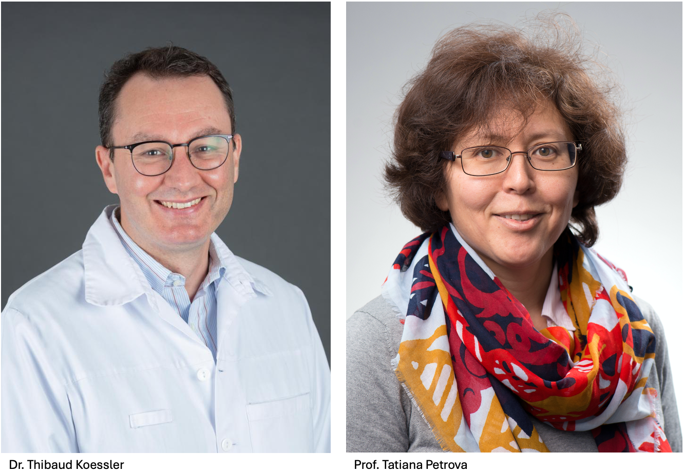
With this TANDEM project, Prof. Petrova and Dr. Koessler will collaborate to investigate the therapeutic and diagnostic potential of these findings in colorectal cancer patients. They aim to do this by analyzing the metabolites that get released in response to chemotherapy, by identifying the liver metastasis niche, and by examining the effect of the metabolites on patient-derived organoids (miniature versions of liver cultivated in vitro).
More specifically, they will:
- Profile the metabolites that are released in response to chemotherapy in CRC patients and relevant animal models.
- Characterize alterations in the liver metastasis niche in response to chemotherapy and gut microbiota components.
- Analyze how the released factors act on the growth of patient-derived organoids and metastasis formation in vivo.
This project will provide insight into how the response of organs to chemotherapy can directly influence patient outcome. The translational goal of this project is to address the current lack of biomarkers that could predict the sensitivity of chemotherapy in clinical practice. Ultimately, this could improve both diagnostic tools and therapeutic options.
Evaluation of the neoantigen-specific T cell response in pleural carcinosis managed by pressurized intrathoracic hyperthermic aerosol cisplatin chemotherapy (PITHAC)
Pleural cancer occurs outside the lungs, in the cavity between the lungs and the chest wall that contains a lubricating fluid, as well as along the pleural lining, a membrane surrounding the lungs and lining the chest cavity. Cancer in the pleural cavity has usually spread from somewhere else in the body, most commonly from lung cancer, but it can also come from the breast, ovary, pancreas, colon, or other locations. Given that pleural tumors are almost always metastatic and difficult to operate, prognosis is poor. One in four patients survives five years after diagnosis of pleural carcinoma. Fortunately, the incidence is low, affecting one in 2’000 cancer patients.
A novel therapeutic approach for pleural carcinoma combines localized, pressurized drug delivery with heat-induced immune stimulation, and goes by the name of PITHAC (pressurized intrapleural hyperthermic aerosol chemotherapy). It is thought that PITHAC triggers a tumor-specific immune response, yet its efficacy in pleural carcinosis remains poorly understood.
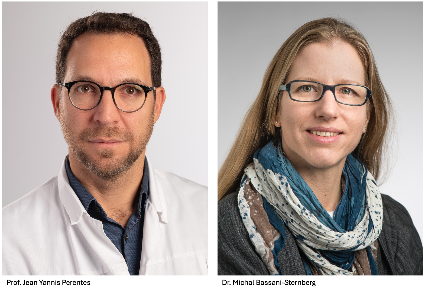
This novel project combines a clinician with expertise in the treatment of pleural carcinosis and a biochemical protein analysis expert. The aim is to determine if PITHAC induces novel antigens on the tumor cells, which in turn trigger a neoantigen-specific response in the immune system, notably in T-cells. By determining the antigenic landscape in the tumors of patients with pleural carcinosis, this study will determine if the mode of action of PITHAC includes the induction of a protective neoantigen-specific T-cell response. If so, it would then be reasonable to combine PITHAC with immunotherapies to make them more efficacious.
The researchers will apply this analysis to the patients enrolled in a Phase I clinical trial that started in 2023 at the CHUV. The trial will assess the feasibility and toxicity of PITHAC in pleural carcinosis patients, and during this trial blood and pleural fluid samples will be collected (after surgery and periodically for a month). The TANDEM grant will finance the analysis of these samples.
The novelty of this project is its longitudinal antigen discovery study, comparing patients before and after therapy, potentially facilitating the integration of PITHAC with immune checkpoint blockade inhibitors and enhancing the study’s translational impact.