Immunotherapy in malignant pleural mesothelioma
Primary pleural cancers such as the malignant pleural mesothelioma, a lung cancer related to asbestos exposure, are cancers that develop in the chest cavity. Mesothelioma is a devastating cancer of high unmet medical need, and its heterogeneous response to immune checkpoint blockade is the rate-limiting factor for improved treatment.
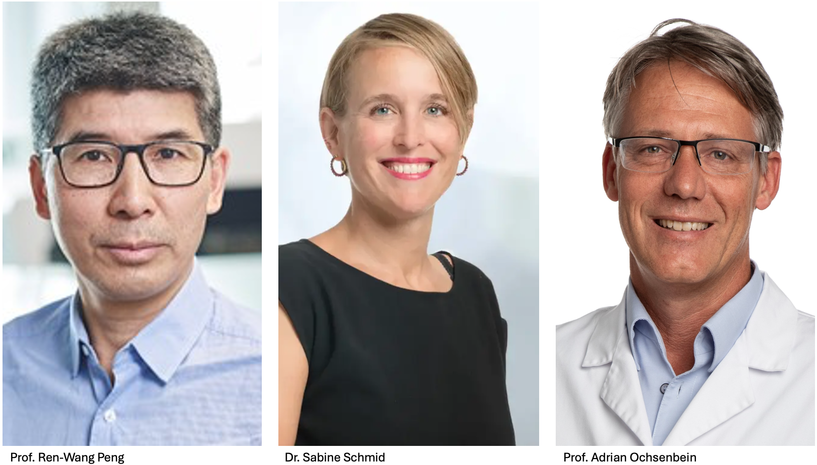
The project led by Profs. Peng and Ochsenbein and Dr. Schmid aims to discover novel immune targets and mechanisms of resistance to immunotherapy in mesothelioma patients. The work builds upon the present state of understanding of mesothelioma treatment, and by determining the variables that correlate with patient response, this study will pave the way for future innovative therapies.
Making use of a large cohort of 109 malignant pleural mesothelioma patients, they will identify the molecular and cellular determinants that correlate with a response to treatments that unblock the patient’s immune response against the tumor. They will use state-of-the-art molecular technologies that visualize the gene expression patterns in the cancer and in the surrounding tissues, cell by cell.
They hope to identify signatures that distinguish responsive from nonresponsive tumors. They will then compare the signature across sections of malignant pleural mesothelioma to determine the effects of the treatments on tumor cell behavior, examining not only the tumor but also the immune cell response to the treatment. To confirm what they will have learned from patient samples, they will then use mouse models of the disease, applying various inhibitors to the mice bearing human mesothelioma. Finally, they will combine immune checkpoint inhibitor therapies with effective inhibitors of growth, both in mouse models and eventually in patients.
The goal is to improve treatments against this devasting disease.
Improving the efficiency of immunotherapy for acute myeloid leukemia
Acute myeloid leukemia (AML) is a cancer that starts in the blood-forming cells of the blood marrow, and which results in the rapid death of the patient if left untreated. Formerly, treatment was based on chemotherapy followed by a hematopoietic stem cell transplantation. More recently, immunotherapy, which programs killer T-cells to attack the cancer cells, has been attempted. Unfortunately, even after intensive and aggressive treatments, a large portion of patients still relapse, and immunotherapy tends to attack healthy as well as leukemic cells. New, innovative treatments and approaches for AML are an urgent unmet clinical need.
To date, the search for AML-specific targets for targeting by cancer-killing CAR-T immune cells, has been unsuccessful. The scientists in this project are now trying a different approach by reversing the therapeutic concept. Their primary goal is now a complete eradication of the disease, including the leukemic stem cells (LSC), while protecting healthy hematopoietic stem cells from the immunotherapeutic attack. This should prevent relapse and overall improve patient outcome.
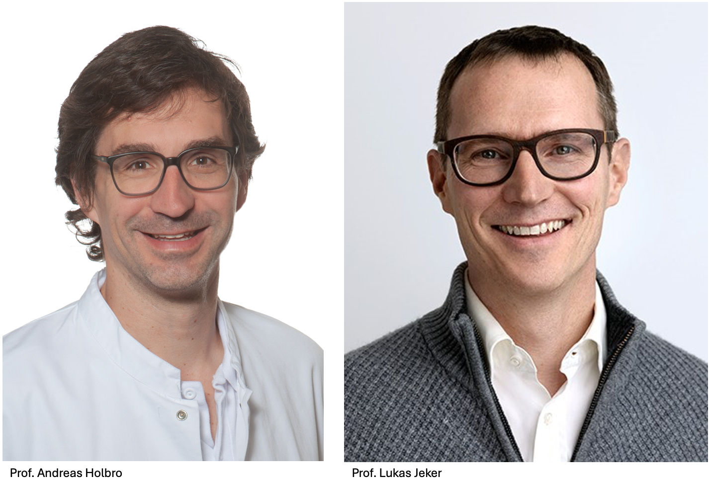
The TANDEM team, consisting of Prof. Jeker and Prof. Holbro, will investigate a potential “cloaking mechanism” that can be used to shield healthy hematopoietic stem cells from the agents that are used to kill the cancer cells. To do this, the scientists need a cell marker (cell surface protein) that is present on both the healthy and the cancer cells, and which they can modify genetically on the hematopoietic stem cells taken from the patient. The protected hematopoietic stem cells will then be re-introduced after AML therapy to regenerate the bone marrow, providing the altered protein does not prevent the cells from functioning normally. The AML-directed immunotherapy will be introduced into the patient and will only recognize the targeted marker on the cancer cells, since the healthy cells carry an altered or mutated marker. This could favor recovery, prevent relapse and improve efficacy of AML treatment.
Immunotherapy for sarcoma treatment

Sarcoma defines an uncommon group of cancers which arise in bones and connective tissue such as fat and muscle. In most cases, it is not clear what causes sarcoma, although family history and exposure to chemicals or radiation may increase risk. There are many types of sarcomas, many being particularly prevalent in pediatric patients, and symptoms depend on tumor type and location.
Current treatments are primarily surgery, radiation, and chemotherapy, yet these offer limited success, especially for advanced cases. There have been some promising results with targeted therapies, but unfortunately, responses are often short-lived. Moreover, immunotherapy, particularly chimeric antigen receptor (CAR) T-cell therapy, presents challenges due to the suppressive tumor microenvironment.
Dr. Digklia and Dr. Irving aim to address the limitations of current sarcoma treatments through a multidisciplinary approach. This will require them to deepen their understanding of sarcoma tumor characteristics, in order to subsequently design and create novel CAR-T cells tailored to sarcoma, and to test ways to enhance their efficacy.
Specifically, this team composed of one clinician and one basic scientist will:
- Characterize sarcoma biopsies from patients at the Sarcoma Center of the CHUV to understand the molecular source of tyrosine kinase inhibitor (TKI) resistance and the barriers to CAR-T cell therapy.
- Develop and engineer new CAR-T cells, targeting specific antigens (B7H3, GD2, and EphA2) for reactivity against sarcoma tissue, based on biopsies and cell lines.
- Optimize combinatorial CAR-T cell therapies for clinical translation, making use of TKIs or other drugs to enhance CAR-T efficacy.
Overall, the goal is to design new, effective treatments for sarcomas, and to test next-generation CAR-T cells that are designed based on their in vitro/in situ results. The scientists will genetically engineer new receptors for CAR-T cells and use their analysis to design new stratification tools for the tumors. Besides CAR-T engineering, they will also analyze the non-cancer tissue surrounding the tumor, to improve the CAR-T cells’ access to the tumor.
The proposed CAR-T targets are well-established and have been used in neuroblastoma, but here the scientists focus on a rare tumor of connective tissue origin, with a persistently bad prognosis. The project is innovative, meets unmet needs and displays a highly collaborative translational aspect with potential to improve the lives of patients living with untreatable sarcoma.
Exploring the anti-tumor functions of immune cells in bladder cancer
Bladder cancer (BCa) is a major health concern, causing approximately 1’400 deaths every year in Switzerland alone. In comparison to prostate cancer, for which recent advances have increased the 5-year survival rates significantly, progress on bladder cancer has stalled. BCa are classified according to how invasive the tumor is at the time of the initial diagnosis. Tumor recurrence is frequent, even when the patient was initially categorized as low risk. For medium- and high-risk patients, it is even worse, as their tumors frequently progress to a muscle-invasive state that requires cystectomy (removal of the bladder). A major challenge is to prevent an initially non-invasive bladder cancer from progressing to muscle invasive disease, which has a much worse prognosis.
So far, the therapeutic approach most commonly used is an intravesical therapy. In this procedure, an immune stimulating agent (such as the Bacillus Calmette-Guerin inoculum (BCG)) is introduced into the bladder to prevent or at least delay tumor recurrence and/or growth. Unfortunately, about 20 to 30% of all patients discontinue the treatment due to severe side effects. Also, it has been shown that even when treated, 20% of all patients experience early recurrence, and only about 45% remain cancer-free for five years. There is an urgent need to find new prognostic tools to identify patients at risk for BCG failure and to predict tumor recurrence/progression. More robust prediction tools may be able to improve the patient’s life quality.
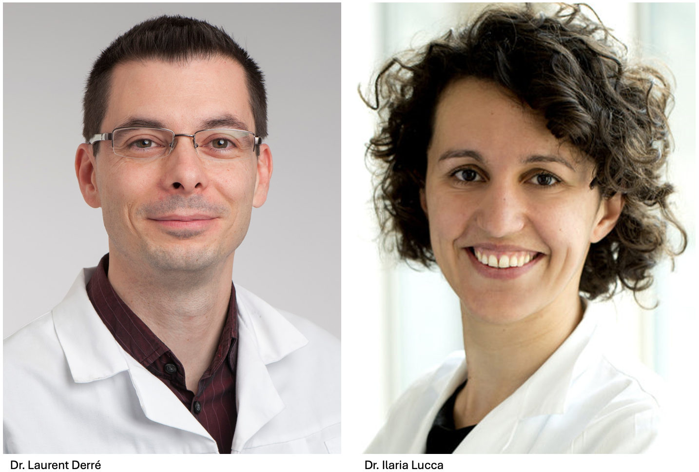
This TANDEM project aims to explore the potential of Vẟ2 T cells (a subtype of T lymphocytes) in this respect. These cells are a subgroup of T cells that infiltrate tumors (called tumor-infiltrating lymphocytes). Recent research has shown that they have the ability to control tumor growth in mice. It remains to be shown that this is relevant to humans, which is why more research using human samples is needed. Specifically, this project will unveil the Vẟ2 T transcriptomic landscape at the single cell level, with the hope that it can serve to identify new biomarkers and allow for the development of new treatments for BCa.
New biomarkers from lymph nodes for programming immunotherapy against triple-negative breast cancer
Breast cancer (BC) remains one of the leading causes of death among women, and 80% of breast cancer deaths are caused by metastatic disease. The majority of these deaths occur in hormone receptor positive patients (HR+), which constitute 80% of all diagnosed breast cancers. In these tumors, the hormone estrogen signals through its receptor to drive tumor growth. Anti-hormone therapy, also called endocrine therapy, which blocks estrogen receptor signalling activity, is the standard treatment for these tumors. Unfortunately, resistance to this therapy increases over time and nearly all HR+ BC patients become refractory to endocrine therapy, generating a large unmet clinical need for new ways to treat HR+ BC patients.
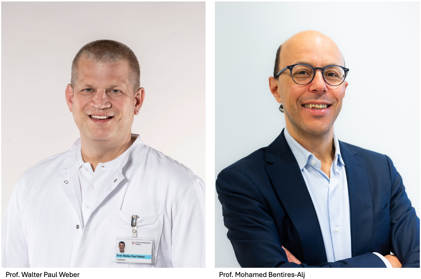
This TANDEM study addresses this urgent unmet need by aiming to identify predictive biomarkers for therapy guidance and patient selection for immunotherapy against HR+ BC. Immunotherapy represents a paradigm shift in the treatment protocols for BC. Yet, to date, it has had little efficacy against recurrent and metastatic BC.
The approaches used in the past to characterize immune therapy-related biomarkers have focused only on the tumor, and no one has checked if promising markers for immune checkpoint therapy can be detected in nearby tissues. This project will investigate the potential of identifying immunological markers in tumor-draining lymph nodes (TDLN). TDLN are the first sites at which the immune system encounters tumor antigens, and these lymph nodes help orchestrate the anti-tumor adaptive immune response. In short, this project is based on the hypothesis that biomarkers found in the TDLN at the early stages of the disease may be more effective for programming and predicting responsive immune cells.
The ultimate aim of the project is to find biomarkers in the lymph nodes near HR+ breast tumors that can indicate sensitivity or resistance to immunotherapy.
Analyzing the role of the tumor microenvironment in platinum drug-resistant ovarian cancer
High grade serous ovarian carcinoma (HGSOC) is the deadliest gynaecological cancer, with a median survival rate of 3 years. Standard treatment involves surgery followed by chemotherapy, typically using platinum and taxane agents. While chemotherapy frequently works well initially to shrink tumors, most women later develop platinum-resistant tumors, that are often fatal. This TANDEM project aims to understand the underlying factor of this resistance, in order to improve immunotherapeutic approaches and develop more effective treatments.
Treatment of HGSOC is a major clinical hurdle, not only because of the commonly developed resistance to chemotherapy, but also due to the fact that it does not respond to recently discovered therapies such as immune checkpoint and targeted T-cell therapies. The reasons for its chemotherapy resistance and insensitivity to immune checkpoint therapy remain unclear.
In this project, the team uses a unique set of patient-derived samples that were collected after resistance had developed and applies cutting-edge molecular technologies to analyze the spatial distribution of cellular and subcellular compartments. Through this, they aim to understand the disease heterogeneity and pinpoint changes that result in resistance.
Preliminary data suggest that platinum-resistant HGSOC is characterized by the accumulation of immune cells called tumor-associated macrophages. These cells are the most common cells in the tumor microenvironment and play a vital role in cancer survival and progression. Leveraging imaging and sequencing tools, the team will identify cell types responsible for resistance, and will gain insight into the spatial distribution and status of the tumor associated macrophages. In this way, they hope to decipher the role these macrophages play in the platinum resistance of HGSOC. Ultimately, these results may lead to an improvement of immunotherapeutic approaches, specifically in the case of platinum-resistant HGSOC.
Identification of new targets to treat prostate cancers that are non-responsive to available treatments
Prostate cancer is the most common cancer among men, affecting 1 in 8, and is a leading cause of cancer-linked mortality and morbidity. Even though localized prostate cancer is highly treatable with surgery, radiation therapy, or active surveillance, survival rates are poor for men with metastatic disease. Existing hormonal therapies in general induce responses initially, but in the majority of cases, their initial effectiveness fades and eventually fails, leading to metastatic castration-resistant prostate cancer (mCRPC).
In addition to hormonal therapy, one of the most promising current treatment regimes for mCRPC uses radioactive compounds linked to antibodies targeting a cell surface protein on mCRPC cancer cells. Such targeted markers are unique to tumor cells, and the binding of high-energy radioactivity, which acts over a small distance, kills these cells efficiently. Unfortunately, up to 30% of patients fail to express the proteins necessary for the radioactive antibody conjugate to recognize the tumor cells and are thus not eligible for this therapy. In addition, only 50% of the patients that express the appropriate marker protein respond well to the treatment.
The main goal of this project is to nominate new therapeutic targets for patients with mCRPC who are not eligible or resistant to this treatment by identifying new surface proteins unique to prostate cancer. This is a laborious process as many tumors need to be sampled and tested to find proteins uniquely expressed on the surface of the cancer cells and not on normal cells. Such probes will ensure that only the tumor cells are being targeted by the radioactive substance during the treatment. This is particularly important as the probe gets introduced into the bloodstream and not injected locally. Not only would this provide treatment for men who at the moment are not eligible for a theranostic treatment, but it could also lead to the further development of other therapy modalities for these patients, using targeted therapies such as CAR-T.
Investigating the connection between circadian system and lung tumor generation to personalize time schedules for chemo-immunotherapy
Lung cancer is one of the most prevalent causes of cancer-related deaths world-wide, largely because most patients have already developed metastases at the time of diagnosis. Besides a need for earlier diagnosis, there is a fundamental lack of effective therapies for lung cancer. As a result, lung cancer has a poor prognosis and low survival rate. In this collaborative TANDEM project, a basic scientist, a surgeon, and an oncologists aim to innovate lung cancer therapy by making use of the body’s internal “circadian” clock to maximize therapeutic impact.
The circadian system evolved in light-sensitive organisms to serve as an intrinsic biological clock with oscillation periods close to 24 hours, in line with geophysical time. It is the molecular time-keeping system operative in most of the cells in the body which drives our physiological activities. Additionally, linked to this is a cell division clock, which drives both normal growth and tumor development. Upon malignant transformation, that is the generation of cancer cells from normal cells, both of the above-mentioned cell-control systems undergo massive changes, resulting in tumor formation.
This project proposes to optimize chronotherapy for lung cancer. Chronotherapy means that the “treatment schedule” is timed in order to align the introduction of medicine with the patient’s natural circadian rhythms. Preliminary data show that for certain types of cancer, the coordination of the administration of anti-tumor drugs at certain times of day improves the effectiveness of chemotherapy and reduces toxicity. Inspired by this emerging potential, the TANDEM team will study the chronobiology of lung cancer, to see if the coordination of therapy with the circadian cycle can improve the outcome for lung cancer patients.
The project has two goals. First, they will refine the diagnostics of lung cancer, and second, they will develop personalised time schedules for the administration of chemo-immunotherapy. This will be achieved by analysing the interaction of the circadian clock with lung cancer progression, and secondly, by analysing the reaction of the patients to therapy administered at different daytimes. This should enable an optimization of lung cancer therapy and improve personalized care.
Using tissue derived from the patient to predict effectiveness of different treatments to find the best one for each patient
The use of molecular and genetic approaches to personalize medical treatments is well on its way to transform cancer therapy. This is because personalized medicine can generate customized therapies and avoid the use of ineffective, and often debilitating, molecules. Currently, cancer treatment is based on tumor stage, mutation profile, and clinical history, while crucial factors such as the tumor heterogeneity and its microenvironment are rarely taken into account. These latter factors, however, are often the most variable and can influence therapy response. Thus, there is an urgent need to incorporate patient-specific data into decisions on the choice of treatment.
This project aims to develop an automated culture system of patient-derived tumor explants. These tumor avatars are unique to individual patients and provide a platform to test sensitivity of each tumour to various treatments. This information could be used to anticipate clinical response, and therefore could guide the hemato-oncologist in selecting the most effective molecule for each patient. In this project the team works with patients affected by non-Hodgkin lymphoma, a group of cancers originating from mature lymphocytes (type of white blood cell).
The team has a number of promising preliminary results. First, the basic research team has developed a method to culture small fragments of the tumor tissue taken from the patient in such a way that key features of the tissue including the cellular composition and architecture are preserved. These fragments, called lymphomoids, can subsequently be used to test the sensitivity to various therapies. Ultimately, the goal is to optimize the lymphomoid technology as a clinical tool to find the most suitable treatment for each lymphoma patient. The team will use cutting-edge image analysis of spatial features to understand the effect of the treatment on both the lymphoma and the neighbouring cells forming the tumor microenvironment. In addition to better tailoring existing treatment to specific patients, this technology can also be used to discover novel therapies.
Ineffective therapies are associated with potential toxicities and ultimately lead to the emergence of resistant diseases that are more difficult to treat. Therefore, implementing a technology that could directly identify these inefficient treatments in routine clinical practice would be groundbreaking and could significantly improve patients’ prognosis and their quality of life.
Analyzing tertiary lymphoid structures as part of the brain tumor environment to develop immune therapies against glioblastoma
Glioblastoma (GBM) is the most common and malignant primary brain tumor in adults. The aggressive and invasive nature of the tumor and its heterogeneity often render it resistant to standard therapies, including chemotherapy, radiation and surgery, leading to a survival rate of less than two years. In this TANDEM collaboration, the team hopes to improve the outcome of GBM treatments by advancing their understanding of the interaction between this tumor and the cellular environment that surrounds it.
Tertiary lymphoid structures (TLS) are ectopic (misplaced) parts of the lymphatic system that develop in non-lymphoid tissues, and which form, importantly, at sites of chronic inflammation such as tumors. Past work has shown that TLS are highly relevant to the prognosis of cancer patients as they form part of the cellular environment that surrounds the tumor, the TME. A major focus of anti-cancer research has been on the macrophages found in TLS, as these white blood cells can either promote or hinder tumor growth, by helping remodel the tissues that surround and support the cancer.
The researchers aim to understand how tertiary lymphoid structures interact with the TME in glioblastoma patients, in order to eventually trigger an anti-tumor immune response in the TLS. Specifically, the project will characterize the repressive TME that blocks normal immune system function, with the ultimate goal of reprogramming the TLS and combining it with CAR-T cell treatment, an advanced T-cell-specific immunotherapy in which T lymphocytes are programmed to recognize tumor cells.
Over the next three years, the team will apply cutting-edge technologies based on the in vivo imaging of gene expression in cells within normal and tumor-containing tissue sections, in order to identify and analyze the contents of the TLS. They aim to understand the intricate interactions of the lymphoid structures with the TME, which helps sustain both the tumor and the TLS. This new knowledge may serve to generate new avenues for therapy, namely the reprogramming of macrophage states in order to support the attack of programmed T cells (CAR-T) on the tumor. The extremely aggressive behavior of glioblastoma and its high mortality rate add urgency to their search for new therapies.