

Analyzing tertiary lymphoid structures as part of the brain tumor environment to develop immune therapies against glioblastoma
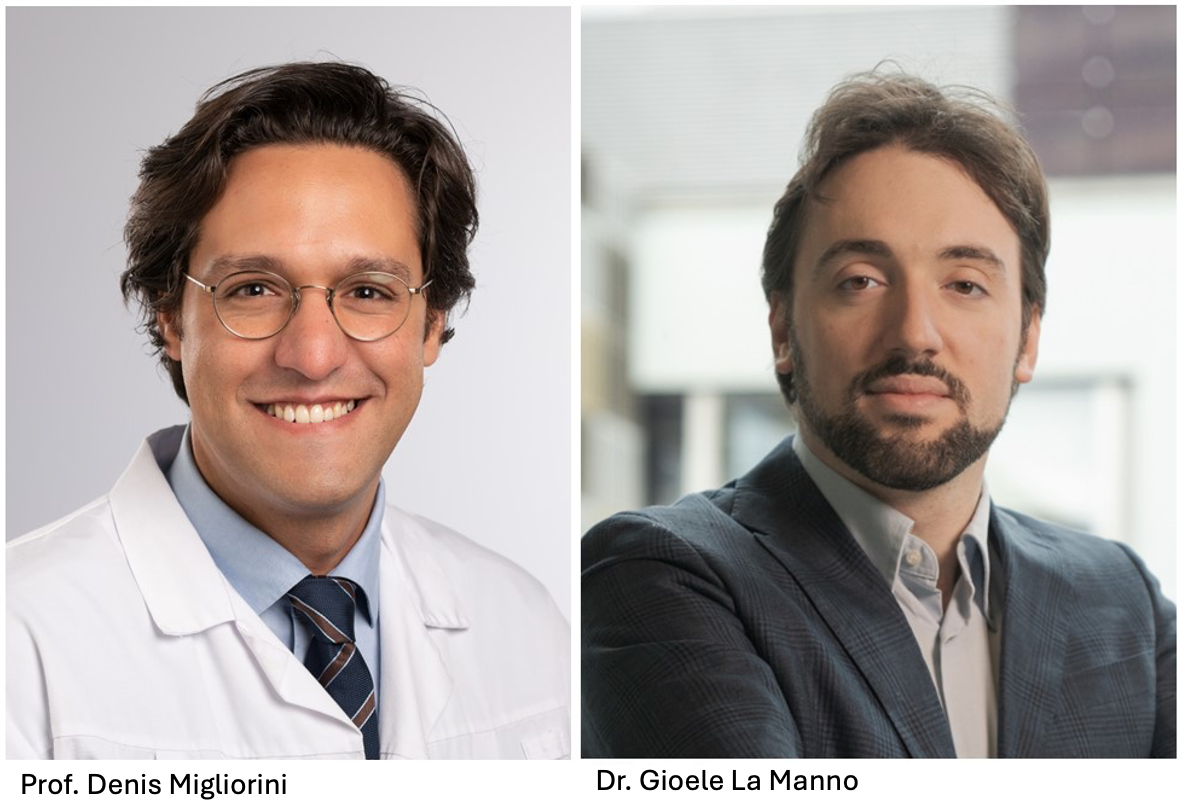
Glioblastoma (GBM) is the most common and malignant primary brain tumor in adults. The aggressive and invasive nature of the tumor and its heterogeneity often render it resistant to standard therapies, including chemotherapy, radiation and surgery, leading to a survival rate of less than two years. In this TANDEM collaboration, the team hopes to improve the outcome of GBM treatments by advancing their understanding of the interaction between this tumor and the cellular environment that surrounds it.
Tertiary lymphoid structures (TLS) are ectopic (misplaced) parts of the lymphatic system that develop in non-lymphoid tissues, and which form, importantly, at sites of chronic inflammation such as tumors. Past work has shown that TLS are highly relevant to the prognosis of cancer patients as they form part of the cellular environment that surrounds the tumor, the TME. A major focus of anti-cancer research has been on the macrophages found in TLS, as these white blood cells can either promote or hinder tumor growth, by helping remodel the tissues that surround and support the cancer.
The researchers aim to understand how tertiary lymphoid structures interact with the TME in glioblastoma patients, in order to eventually trigger an anti-tumor immune response in the TLS. Specifically, the project will characterize the repressive TME that blocks normal immune system function, with the ultimate goal of reprogramming the TLS and combining it with CAR-T cell treatment, an advanced T-cell-specific immunotherapy in which T lymphocytes are programmed to recognize tumor cells.
Over the next three years, the team will apply cutting-edge technologies based on the in vivo imaging of gene expression in cells within normal and tumor-containing tissue sections, in order to identify and analyze the contents of the TLS. They aim to understand the intricate interactions of the lymphoid structures with the TME, which helps sustain both the tumor and the TLS. This new knowledge may serve to generate new avenues for therapy, namely the reprogramming of macrophage states in order to support the attack of programmed T cells (CAR-T) on the tumor. The extremely aggressive behavior of glioblastoma and its high mortality rate add urgency to their search for new therapies.
Development of an endoscope to better define tumor margins during surgery
Supported with the generous contribution of the LARDECO Foundation
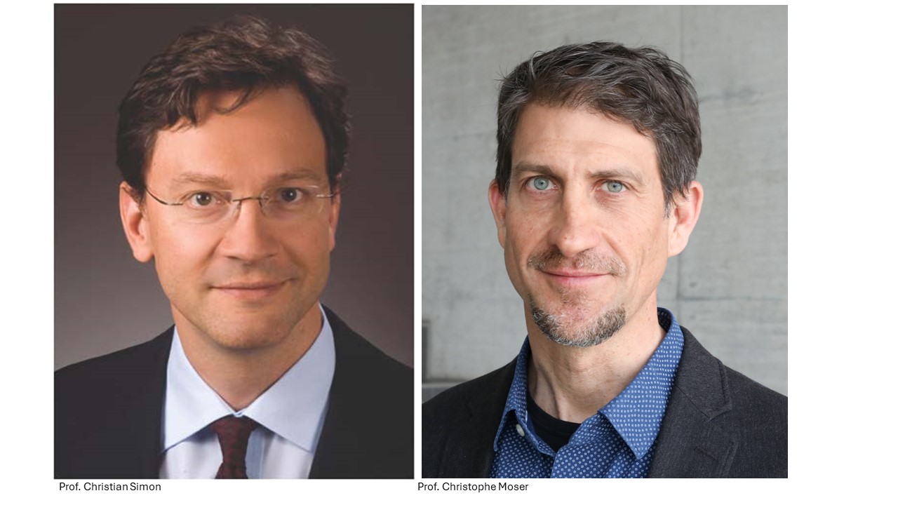
Neck and head cancers (HNC) are lethal and mutilating. With over 150’000 new cases diagnosed each year in Europe alone and 370’000 deaths world-wide, these cancers have a significant impact on the human population. The main issue with HNC is that they have characteristic infiltrative growth, which means that the disease can escape eradication by local surgery and spread. This TANDEM project aims to improve the technology used to make HNC surgery more efficient.
For more than 50% of the HNC patients the first-line treatment is surgery. During those interventions it is essential that the surgical margin (the “border” between the tumor tissue and healthy tissue) is negative for cancer cells. This requires the excision of the cancer such that even on the microscopic level no tumor cells are left behind. Residual disease can lead to local reoccurrence and death of the patient.
The routinely used surgical techniques have limited resolution and surgeons often have poor visibility of the extension of the tumor, which leads to diseased cells around the edge not being detected. So, even though the surgery is deemed successful, in about 20% of the patients, it is not. Consequently, such patients must undergo further treatments such as chemical and radiation therapy which are aggressive and seriously impact the patient’s quality of life.
This collaboration between clinicians and engineers aims to use recently developed ultra-thin endoscopes – which are minimally invasive due to their small size (thin as a hair!) while still providing high resolution images –that will enable a more precise visualization of tumor cells in situ. Importantly, this technology will be implemented in real time during surgery to enable the surgeon to predict with much higher accuracy where the tumor tissue ends and the healthy tissue begins. Ultimately this will improve the reliability of diagnostics and the rate of success of HNC surgery for these cancer patients.
Expanding the knowledge on the potential of cancer vaccines
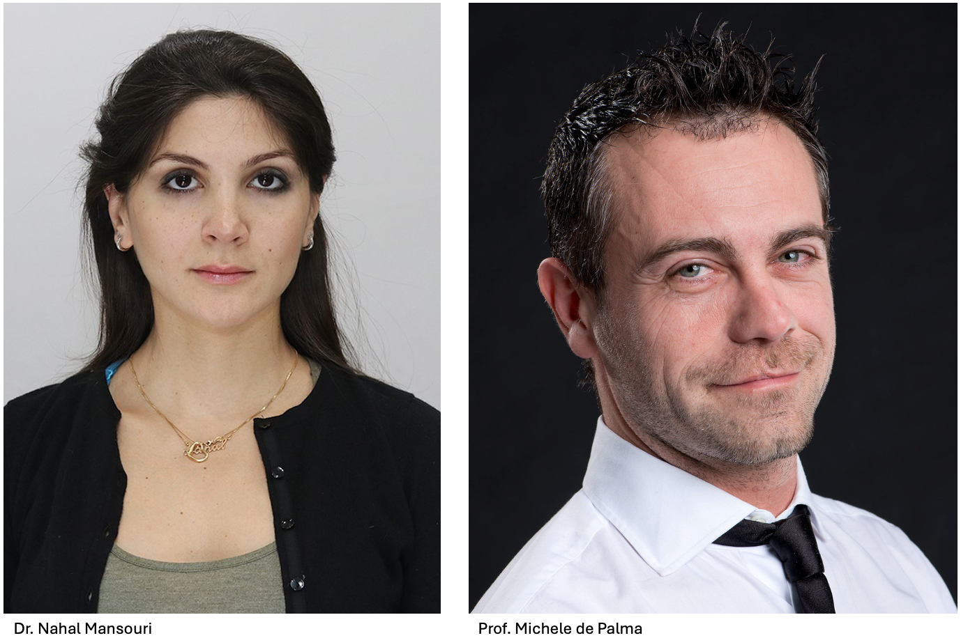
Lung cancer is, to this day, the leading cause of cancer-associated deaths worldwide. There is a dire need for the development of more effective therapies, as lung tumors often become resistant to both conventional and targeted therapies, such as immunotherapies. This collaboration will work on advancing a promising cancer therapy referred to as dendritic cell (DC) vaccines.
DCs play a fundamental role in orchestrating the functions of our immune system. They present antigens on their surface that are recognized by other players in the immune response. Their biological role has long been exploited to develop DC vaccines for patients with cancer. The goal of a DC vaccine is to make the patient’s own immune system recognize and eliminate the cancer cells. More specifically, immature DCs are isolated from a patient with cancer and then exposed to tumor-associated antigens. After reaching full maturity, the cells are reintroduced in the patient to trigger an anti-tumor response. However, this traditional approach has several limitations and has yielded mixed clinical results.
This TANDEM project aims to advance the design of DC vaccines for lung cancer therapy. It exploits a novel type of DC, which is engineered in the laboratory to improve its ability to present tumor antigens to the immune system. This work is poised to improve the therapeutic potential of DC vaccines and will hopefully provide a new treatment strategy for lung cancer patients.
Understanding disease progression in lung cancer
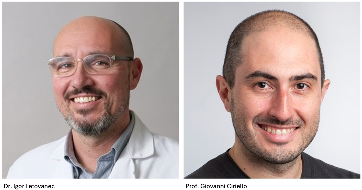
Lung adenocarcinoma (LUAD) represents 40% of all lung cancers, which makes it one of the most common lung cancers. Previous studies have used histopathology (the study of changes in tissues caused by disease) to diagnose and study diseased lung tissue at the microscopic level. This classic approach identified changes in cell morphology and growth patterns that accompany disease progression. The overarching aim of the current collaboration is to use new techniques to predict more accurately how the cancer will progress and whether or not it will react to treatment.
Disease progression is driven by the plasticity of cell identity and a coincident reshaping of the tumor environment, such that reprogramming is maintained. Based on histopathological analysis, there are four recurrent tumor progression patterns that reflect both tumor aggressivity and patient survival prognosis. The milestones are readily identified and provide a lot of information on disease progression and tumor heterogeneity, both within one patient and among multiple patients:
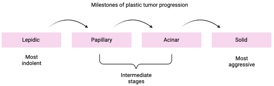
Whereas it is clinically relevant to identify the stages of tumor progression by histopathology, the molecular drivers of the transitions from one state to the next are also crucial. Towards this end, the team has characterized molecularly the transitions of cancer from lepidic to solid tumors, using a combination of techniques for single cell analysis of cancer cells and their interaction with the tumor microenvironment.
The team has the following aims: first is to detect molecular features of the transitions from lepidic to solid tumors across patients (see figure). Secondly, they will deepen our understanding of how tumor progression can be predicted based on the interaction of cancer cells with the tumor cell environment. The overarching goal of the project is to provide new insights into the role of cancer cell plasticity in disease progression and to explore if this helps predict disease progression in individual patients. Ultimately, the results will pave the way for new practices in clinical diagnostics as well as new approaches to lung cancer therapy.